

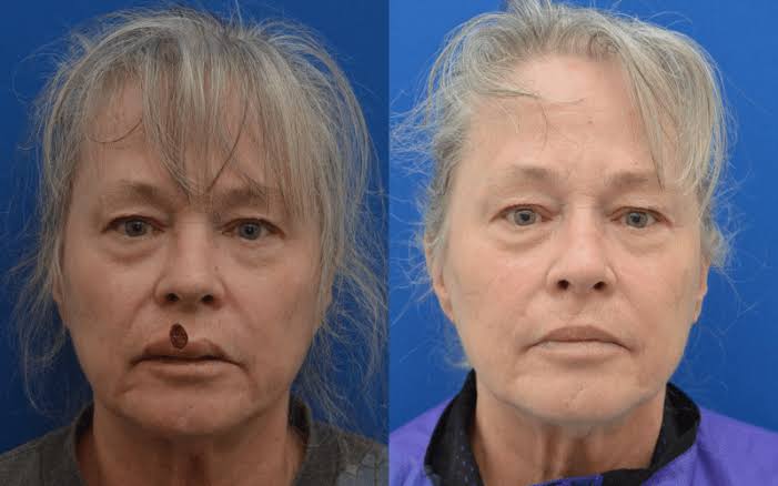
Mohs surgery is a staged technique for removing certain skin cancers, and this includes selected cases of melanoma. The procedure removes thin layers of tissue and examines each layer under a microscope during the same visit. When melanoma is on areas such as the face, this level of precision shapes both treatment planning and reconstruction. Here is more information on the role of this surgery for melanoma:
Addressing Early-stage Melanoma
Early-stage melanoma sometimes responds well to Mohs surgery when the lesion remains confined to the top layers of skin. The dermatologist first confirms the diagnosis and depth with a biopsy, and that information guides whether this technique fits the case. When margins are unclear, a layer-by-layer method helps track stray cells that extend beyond what the eye detects.
The decision to use Mohs surgery typically depends on the location, and the doctor examines the subtype and thickness of the melanoma. Areas with limited spare skin, such as the nose or eyelids, benefit from a method that removes less tissue. When the tumor is still thin and localized, the goal is complete removal while limiting functional changes.
Removing Layers of Tissue
During Mohs surgery, the surgeon removes tissue in stages and studies each stage under a microscope before moving on. This process continues until no melanoma cells appear in the examined sections, and the mapped diagram shows clear margins. The surgeon adjusts the next layer precisely to the areas where cells remain. Surgery involves these steps:
- The visible tumor and a narrow rim of skin are removed first.
- The tissue is marked, processed, and examined under the microscope.
- The map links what the surgeon sees on the slide to the spot on the skin.
- The cycle repeats until the microscopic edges appear free of tumor.
Sparing Healthy Tissue
One advantage of Mohs surgery lies in its focus on preserving healthy tissue while clearing the melanoma. Each stage removes only a thin layer, and the final defect typically stays smaller than with a wide excision. That difference matters in areas like the nose, lips, or eyelids. When less tissue leaves the site, reconstructive options after the procedure sometimes remain more flexible.
Traditional excisions rely on predetermined margins, and those margins occasionally remove more normal skin than necessary. The Mohs mapping process helps the surgeon avoid guessing about where the tumor ends. With a staged approach, the surgeon tailors the next pass to the exact spot where cells still appear.
Since healthy tissue is spared, the cosmetic outcome typically aligns better with surrounding features. It stays closer to baseline, and patients who work in public-facing roles or who rely on precise facial movement sometimes value that balance. The approach also supports careful planning for closures, flaps, or grafts once the site tests clear.
Excluding Advanced Melanoma
Advanced melanoma typically requires a different strategy, so Mohs surgery often plays no role once the tumor invades deeper structures or spreads. When melanoma reaches lymph nodes or distant organs, treatment typically shifts toward systemic therapies and wider surgical approaches. Even for thicker primary tumors without spread, standard excision with broader margins is typically preferred.
Book a Mohs Surgery Consultation
A detailed consultation helps determine whether Mohs surgery fits a specific melanoma diagnosis and location. During that visit, the dermatologist reviews pathology reports, examines the area, and explains alternative treatment paths. Each case differs, so direct discussion with a trained skin cancer specialist offers the most relevant guidance. When melanoma or another skin cancer enters the picture, timely evaluation sets the stage for appropriate treatment.