

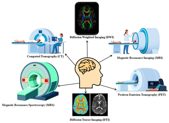
Neuroradiology is a subspecialty focused on imaging the brain, spine, head, and neck. Neuroradiologists interpret imaging studies of the nervous system, supporting care teams that evaluate a wide range of conditions, from stroke and vascular problems to brain tumors and spinal abnormalities. After a traumatic injury, imaging helps physicians identify the location and extent of damage. This information may guide decisions about further testing, monitoring, or treatment. Here is information on neuroradiology diagnostic tests and how they help doctors understand traumatic injuries to the head and spine:
A Myelography
Myelography is a neuroradiology imaging test that examines the spinal canal, spinal cord, and nerve roots. During the procedure, a physician injects contrast material into the fluid-filled space around the spinal cord, making these structures more visible on X-ray or CT images. A neuroradiologist then reviews the images for changes in the shape or position of the spinal cord and nerves. A traumatic event, such as a fall or vehicle collision, may cause spinal damage that is difficult to see with other methods. Myelography helps show areas where the spinal canal has narrowed or where nerve roots may be compressed. Physicians may recommend this test when a patient cannot undergo an MRI, like when someone has implanted medical devices.
A Cerebral Angiography
Another screening option is a cerebral angiography, which produces detailed images of the blood vessels in and around the brain. For this procedure, a physician inserts a thin, flexible tube called a catheter into an artery, usually in the groin or wrist. The catheter is guided through the blood vessels to the arteries of the neck and brain. Contrast material is then injected through the catheter while X-ray images are captured.
Trauma to the head or neck can affect blood vessels as well as surrounding tissue. Cerebral angiography helps physicians identify injuries such as:
- Tears in an Artery Wall
- Abnormal Connections Between Vessels
- Areas of Bleeding
Because the images show blood flow in real time, physicians can observe how blood moves through specific vessels.
An MRI or CT Scan
Magnetic resonance imaging (MRI) and computed tomography (CT) are two of the most common imaging methods in neuroradiology, and each has distinct advantages for evaluating traumatic injuries. MRI uses a strong magnetic field and radio waves to create detailed soft-tissue images, so it doesn’t use ionizing radiation. MRIs can produce clear views of the brain, spinal cord, ligaments, and discs. CT scans use a series of X-ray images taken from different angles, and a computer combines them into cross-sectional views of the head
These images help reveal subtle injuries, such as small areas of bleeding or damage to nerve fibers, that may not appear on other scans. An MRI exam often takes 30 to 60 minutes, but patients must remain still inside the scanner throughout the scan. Head CT scans typically take only a few minutes; this speed makes them a common imaging test in emergency settings after a head injury. CT images can show skull fractures, bleeding within the brain, and swelling. Physicians may use CT results to decide whether additional imaging, including MRI, is needed.
Learn More About Neuroradiology
Neuroradiology gives physicians several tools to examine the brain, spine, and blood vessels after trauma. Myelography shows the spinal canal and nerve roots, while cerebral angiography shows the blood vessels of the brain and neck. MRI and CT scans each produce images that serve different purposes during evaluation. If you have questions about an imaging test, speak with your physician or radiologist. They can explain how a specific test works and what to expect before, during, and after the procedure.